FREE WEBINAR

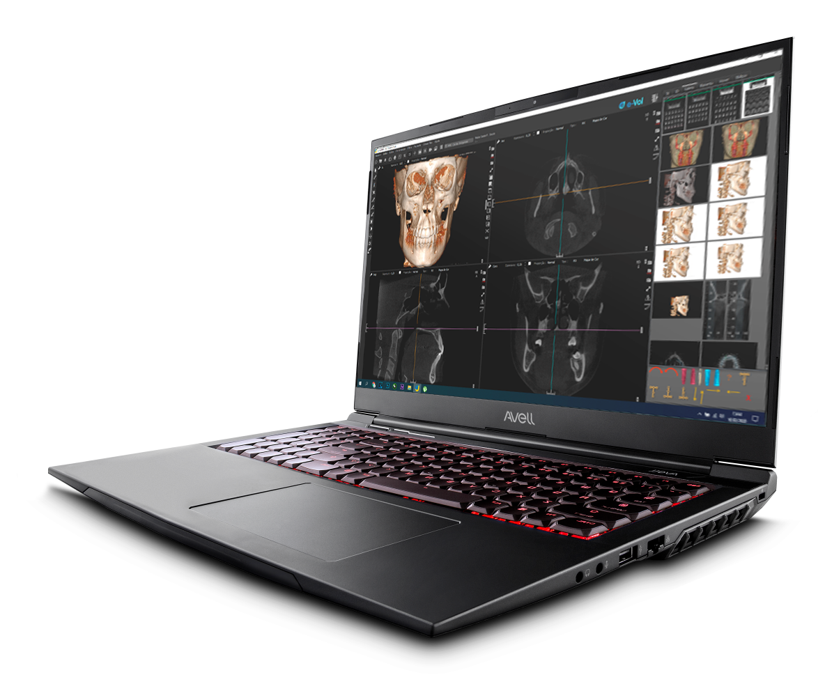
The eVol DXS is the definitive CBCT software to enhance imaging and improve imaging diagnosis. Its unique Task-Specific Rendering (TSR) filters and features allow for the ultimate control of the CBCT data leading to sharper images and a greater display of detail:
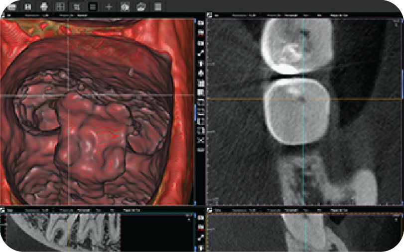
Pulp Horn Filter
Allows a detailed, three-dimensional view of the structure
of the pulp horn and the surrounding area
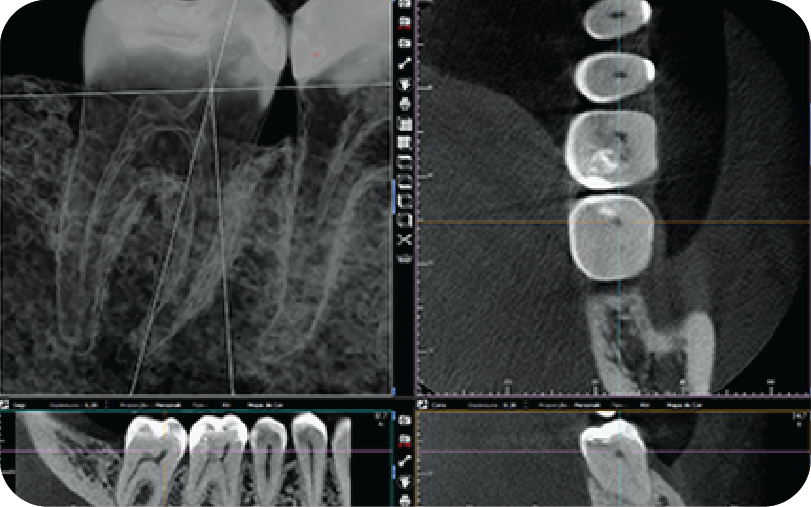
Dental Pulp Filter
Allows the visualization of the shape and size
of the pulp and pulp horns
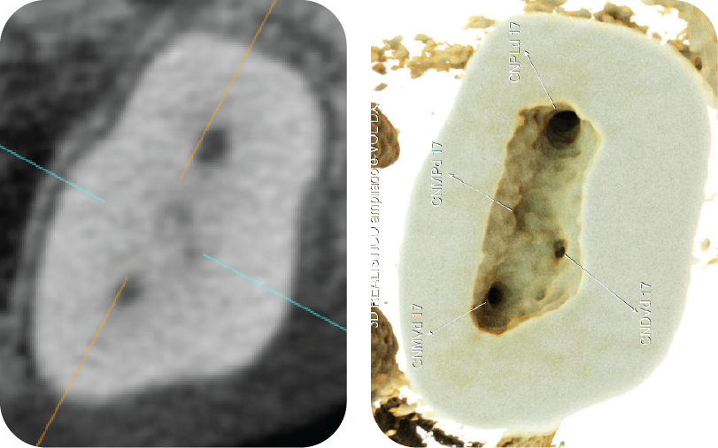
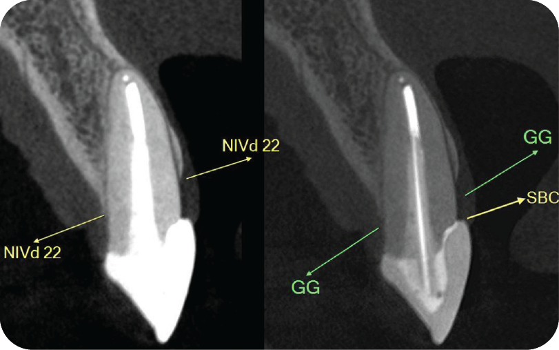
ACI Filter Realistic
Identify accessory
canals with ease
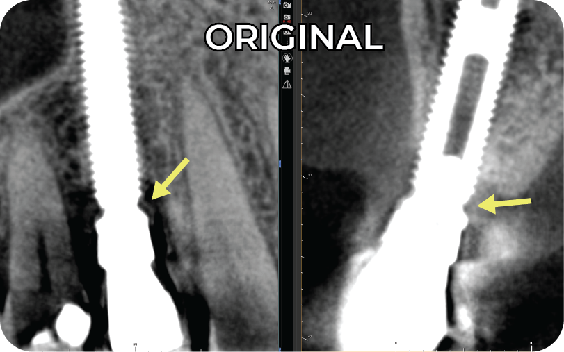
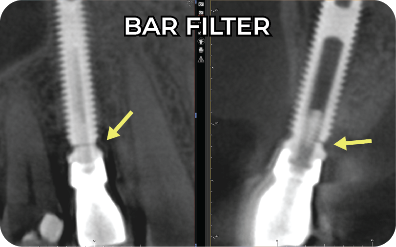
BAR Blooming Artifact Reduction Filter
Minimizes white contrast artifacts generated by metallic and dense materials
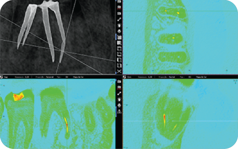
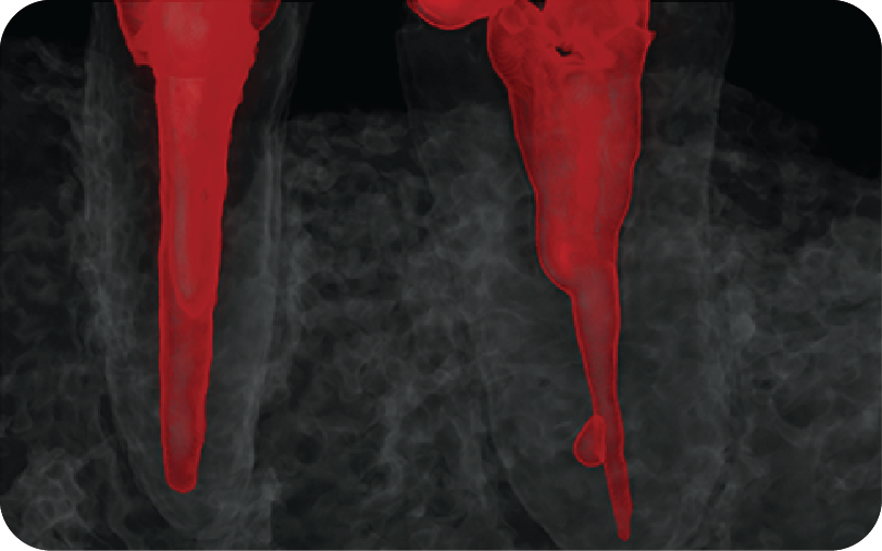
Fractured Instrument Filter
Helps identify a fractured instrument
inside a filled canal
Endodontic Fillings Filter
Helps differentiate different
types of filling materials
LESS TIME WITH TOMOGRAPHIES
improve your endodontic diagnosis
With e-Vol you spend less time browsing a tomography Faster diagnosis
Spend less time interpreting images
Protect your investment
Extract as much information from your CBCT scanner
WOW your patients
Use the most powerful 3D rendering tools powered by AI to mesmerize your patients and better explain treatment plans
Elevate your diagnostic skills
The e-Vol DXS allows you to see the invisible so you can do the impossible
